Cataract, Cataract Surgery & Clear Lens Extraction
(白内障手术)
What is cataract?
A cataract is a common eye condition where the natural lens of the eye becomes cloudy, leading to blurred vision, glare, difficulty in reading, driving and walking around. Cataracts often develop gradually with age but it may occur at birth (i.e. congenital cataract) or can result from eye injuries, medical conditions such as diabetes, or use of steroids.
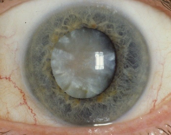
Photo credit: National Eye Institute
Symptoms of cataract
- Blurry or cloudy vision
- Increased sensitivity to bright lights and glare
- Reduced contrast
- Difficulty in reading or seeing in dim light conditions
- Difficulty in driving
- Frequent changes in glasses prescription without much improvement
If left untreated, cataracts can significantly affect daily activities, making it harder to drive, read, or recognise faces.
Treatment
Cataract surgery is a safe and effective procedure to restore clear vision. It involves removing the cloudy lens and replacing it with an artificial intraocular lens (IOL). Surgery is usually recommended when cataract starts interfering with your daily activities.
Preoperative assessment
Before cataract surgery, a thorough preoperative assessment is conducted to ensure optimal surgical outcomes. This includes:
- Slit-Lamp Examination: A detailed evaluation of the eye using a high-magnification microscope to assess the severity of the cataract and check for other eye conditions such as corneal disease, glaucoma, or retinal disorders.
- Tear Film Assessment: Evaluates dry eye status as dry eyes can impact surgical accuracy, choice of IOL, and postoperative comfort.
- Biometry: Measurements of the eye’s length and curvature to determine the appropriate intraocular lens (IOL) power. This is typically performed using optical coherence biometry or ultrasound.
- Corneal Topography: Assesses the shape and curvature of the cornea, which is especially important for patients considering toric or multifocal IOLs.
- Pupil Dilation: The pupils are dilated to allow a detailed view of the retina and optic nerve, ensuring there are no underlying conditions that may affect visual recovery.
- Discussion of IOL Options: Based on lifestyle needs, visual goals, and ocular measurements, the most suitable intraocular lens (IOL) type is selected.
Types of intraocular lens (IOL)
There are different types of IOLs to suit individual needs:
- Monofocal IOLs: Provide clear vision at a single distance (near or far), often requiring glasses for certain tasks. This is usually the type of IOL implanted in NHS settings.
- Multifocal IOLs: Designed to provide clear vision at multiple distances (including distance, intermediate and near), reducing dependence on glasses. However, they may cause glare of halos in some patients. This type of lens is usually available in the private sector only.
- Extended Depth of Focus (EDOF) IOLs: Provide a continuous range of vision from distance to intermediate and/or near, reducing the need for glasses while maintaining contrast and clarity. However, they may cause glare of halos in some patients (but less common than the multifocal IOLs). This type of lens is usually available in the private sector only.
- Toric IOLs: Correct corneal astigmatism for clearer vision. This may be available as monofocal toric, multifocal toric and EDOF toric IOL. The multifocal and EDOF toric lenses are usually available in the private sector only.
Choosing the right IOL depends on factors such as lifestyle, visual needs, and pre-existing eye conditions. We will discuss the best options for you during the consultation.
The procedure
- Anaesthesia: The procedure is usually performed under local anaesthetic (>95%), meaning you remain awake but feel no pain. However, general anaesthesia may be required occasionally due to patient preference or certain underlying conditions.
- Start of procedure: A sterile drape will be placed over the face. Then, a lid speculum will be inserted to help hold the eye open.
- Lens Removal: A small incision is made in the cornea to allow access to the lens. The cloudy lens is then broken up using ultrasound energy and gently removed from the eye (known as phacoemulsification).
- Lens Replacement: A new intraocular lens (IOL) is inserted to restore focus and clarity.
- Recovery: The procedure typically takes 10-15 minutes, and most patients go home the same day. Vision improves gradually over the following days to weeks.
Potential risks
Cataract surgery is generally a very safe and effective procedure, with 95% of the patients noticing visual improvement after the surgery. However, like any other procedure, cataract surgery is associated with certain risks. These include:
During the operation
- Posterior capsular rupture (PCR): An uncommon but recognised risk (~1%) of cataract surgery. The posterior capsule is a thin membrane that holds the lens in place. During surgery, the surgeon removes the cloudy cataract and leaves the posterior capsule intact to support the implantation of the artificial IOL. Occurrence of PCR may warrant additional procedure such as anterior vitrectomy (removal of the gel of the eye).
- Dropped Nucleus: In certain complicated cases, part of the cataract lens (the nucleus) may drop into the vitreous body, requiring additional surgery.
- Injury to cornea: Uncommonly, the cornea may be affected during the surgery, usually in patients with a small eye (e.g. longsightedness)
- Injury to iris: Uncommonly, the iris may be damaged during the surgery, usually in patients with a small pupil or floppy iris.
After the operation
- Inflammation: Mild inflammation is common after surgery, but severe inflammation (uveitis) can occur in some cases, potentially affecting the recovery process.
- Infection (Endophthalmitis): A rare but serious complication that can lead to severe or complete vision loss (less than 1:1000 risk) if not treated promptly.
- Retinal Detachment: This is a rare complication, but the risk increases in patients who are highly shortsighted or have had previous eye surgeries.
- Posterior Capsule Opacification (PCO): Sometimes referred to as a "secondary cataract," this is a clouding of the lens capsule after surgery, which can cause blurred vision. It can be treated with a simple laser procedure called YAG capsulotomy.
- Vision Problems: Though rare, some patients may experience problems with glare, halos, or double vision after surgery.
- Increased Eye Pressure (leading to glaucoma): A temporary increase in eye pressure can occur, which may require medication to control.
- Corneal swelling / oedema: Swelling of the cornea can lead to blurry vision or discomfort but usually resolves with time, unless the patients have pre-existing corneal weakness such as Fuchs endothelial corneal dystrophy.
- Macular swelling / oedema: Swelling of the macula (the central part of the retina responsible for sharp vision), which usually responds to medical treatment.
What is clear lens extraction?
Clear Lens Extraction (CLE), also known as Refractive Lens Exchange (RLE), is a surgical procedure that removes and replaces natural, clear crystalline lens of the eye with an artificial IOL. Unlike cataract surgery, which is performed to remove a cloudy lens, CLE is done primarily to correct refractive errors such as severe myopia (shortsightedness, "近视"), hyperopia (longsightedness, "远视"), and presbyopia (age-related loss of near vision, "老花"). The surgical procedure and aftercare is the same as the cataract surgery.

© Copyright. All rights reserved.
We need your consent to load the translations
We use a third-party service to translate the website content that may collect data about your activity. Please review the details in the privacy policy and accept the service to view the translations.